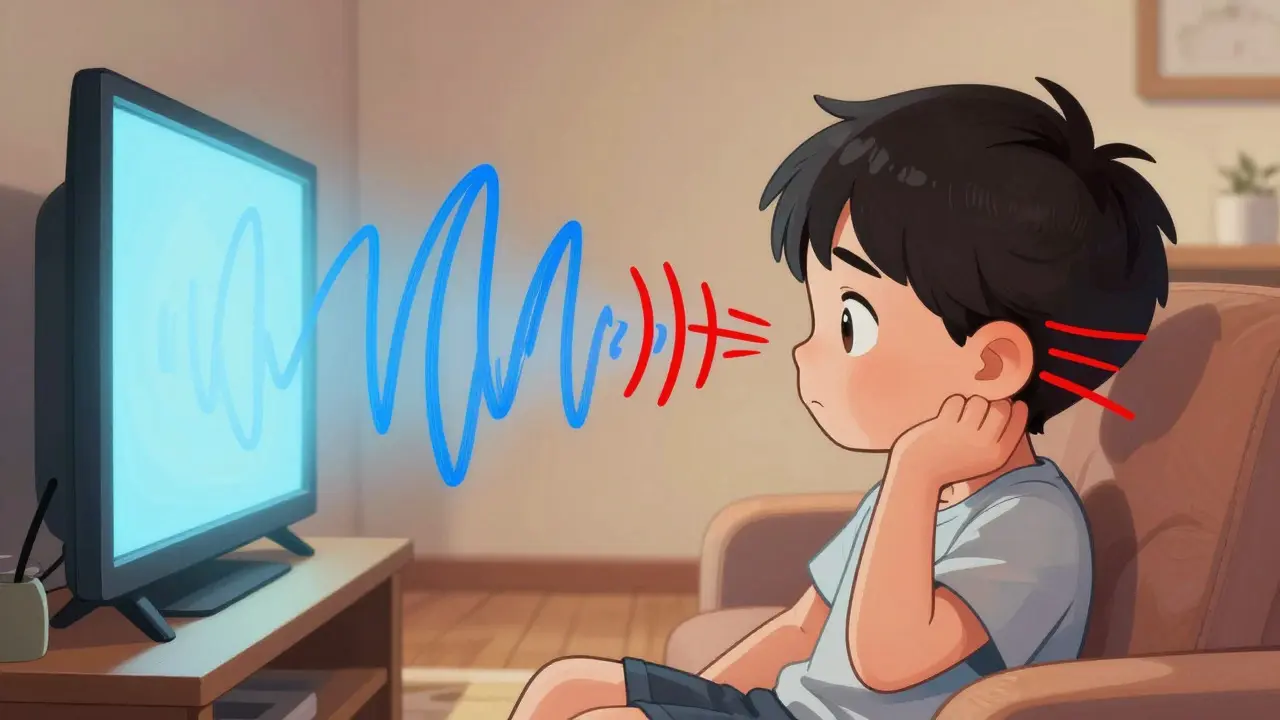
Imagine trying to hold a conversation in a noisy room, but every word sounds like it’s coming from underwater. That muffled, distant quality is often the hallmark of conductive hearing loss, a condition where sound waves struggle to travel through your outer or middle ear to reach the inner ear. Unlike damage to the nerve itself, this type of hearing impairment is usually mechanical-a blockage, a fluid buildup, or a broken bone chain-and that’s the good news: it’s frequently fixable.
You don’t have to live with a plugged-up world just because you’ve had a few bad colds or an old injury. Whether you’re a parent watching your child tilt their head during movie night or an adult noticing that the TV volume keeps creeping up, understanding what’s happening inside your middle ear is the first step toward getting your clarity back. This guide breaks down the common culprits behind these issues and walks you through the surgical repairs that can restore normal hearing without the guesswork.
What Is Conductive Hearing Loss and Why Does It Matter?
Conductive hearing loss occurs when sound cannot properly travel from the outer or middle ear to the cochlea in the inner ear, resulting in reduced sound transmission. Think of your ear as a sophisticated microphone system. The outer ear catches the sound, the eardrum vibrates, and three tiny bones (the ossicles) amplify those vibrations before sending them into the fluid-filled inner ear. If any part of this chain gets jammed, broken, or blocked, the signal weakens before it even reaches the hearing center.
This differs significantly from sensorineural hearing loss, which involves damage to the hair cells in the inner ear or the auditory nerve. With conductive loss, the "hardware" is intact; it’s just not moving freely. According to the American Speech-Language-Hearing Association (ASHA), people with this condition often find soft sounds difficult to hear, while louder noises may seem muffled rather than distorted. It can develop gradually over months or appear suddenly after an injury or severe infection. Sudden onset should always be treated as a medical priority to prevent permanent structural damage.
| Feature | Conductive Hearing Loss | Sensorineural Hearing Loss |
|---|---|---|
| Location of Problem | Outer or Middle Ear | Inner Ear or Auditory Nerve |
| Common Causes | Fluid, wax, perforation, otosclerosis | Aging, noise exposure, genetics |
| Reversibility | Often reversible with treatment/surgery | Usually permanent (managed with aids) |
| Sound Perception | Muffled, low volume | Faint, distorted, high-frequency loss |
Common Middle Ear Problems Causing Hearing Loss
Not all middle ear issues are created equal. Some are temporary annoyances, while others require urgent intervention. Here are the most frequent offenders:
- Otitis Media with Effusion (Glue Ear): This is the most common cause of acquired hearing loss in children. Fluid builds up behind the eardrum without active infection, creating a sticky barrier that dampens sound. Up to 80% of children experience at least one episode by age three.
- Otosclerosis: An inherited condition where the stapes bone (one of the three tiny ear bones) fuses with the surrounding structures. This prevents the bone from vibrating properly, leading to progressive hearing loss. It typically affects adults between ages 30 and 50.
- Tympanic Membrane Perforations: A hole in the eardrum caused by trauma, barotrauma (like flying or diving), or chronic infection. These account for 15-20% of conductive hearing loss cases in adults.
- Cholesteatoma: A non-cancerous growth of skin cells in the middle ear that can erode bone if left untreated. While rare, it requires surgical removal to protect hearing and balance.
- Aural Atresia: A congenital condition where the ear canal fails to develop fully, affecting about 1 in 10,000 births. Sound enters through the skull instead of the canal, causing significant attenuation.
Dr. Aaron Pearlman, Director of Otology at Mass Eye and Ear, notes that determining the exact cause is critical because treatment varies dramatically. Wax impaction needs cleaning; a perforation needs patching; otosclerosis needs bone replacement. Misdiagnosis can lead to unnecessary procedures or missed opportunities for simple fixes.
How Doctors Diagnose Middle Ear Issues
Diagnosing conductive hearing loss isn't just about plugging in headphones at a pharmacy. ASHA emphasizes that proper diagnosis requires the diagnostic skills of an audiologist, not a basic screening test. The standard pathway involves two main components: physical examination and specialized testing.
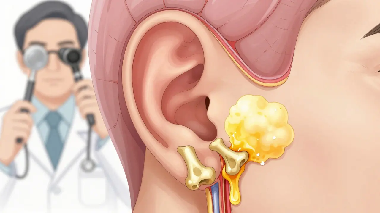
- Otoscopic Examination: Your doctor uses a lighted instrument to look directly at your eardrum. They check for fluid levels, color changes, perforations, or signs of infection. This gives immediate clues about the source of the problem.
- Audiometric Testing: You’ll undergo air and bone conduction tests. Air conduction measures how sound travels through the outer and middle ear, while bone conduction bypasses the middle ear entirely to test the inner ear. A gap between these two results (called an air-bone gap) confirms conductive loss. Gaps typically range from 15 to 60 dB depending on severity.
- Tympanometry: This test measures how well your eardrum moves. A Type B tympanogram indicates fluid presence in 92% of cases of otitis media with effusion, helping doctors confirm if glue ear is the culprit.
The typical diagnostic process takes 2-3 appointments spanning 2-4 weeks. This timeline allows doctors to rule out temporary conditions like a passing cold before considering long-term solutions. For surgical candidates, a high-resolution CT scan of the temporal bone is often required to map the anatomy precisely, ensuring the surgeon knows exactly what they’re working with.
Surgical Repair Options for Different Conditions
When medical management fails-usually defined as hearing loss exceeding 25-30 dB persisting for 3-4 months-surgery becomes the next logical step. The procedure chosen depends entirely on the specific pathology identified during diagnosis.
Tympanoplasty for Eardrum Perforations
If your eardrum has a hole, a tympanoplasty is the standard fix. Surgeons use a graft, often taken from your own tissue (temporalis fascia) or bioengineered material, to patch the tear. Success rates are high: 85-95% for small perforations and 70-85% for larger ones. Postoperatively, you’ll need to avoid water exposure for six weeks and pressure changes (like flying) for eight weeks to allow the graft to heal securely.
Stapedectomy for Otosclerosis
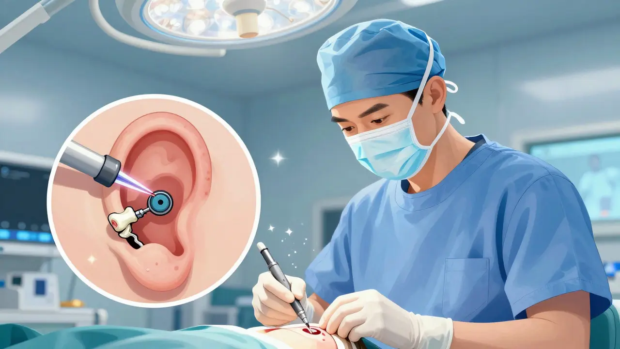
For otosclerosis, the fused stapes bone is replaced with a prosthetic device. Modern laser-assisted techniques have revolutionized this procedure, reducing complication rates from 15% historically to less than 2%. In 80-90% of cases, the air-bone gap closes to within 10 dB post-surgery, meaning most patients regain near-normal hearing. Patients often report being able to hear whispers again and no longer needing to turn up the television volume.
Myringotomy and Tubes for Glue Ear
In children with persistent fluid, myringotomy involves making a tiny incision in the eardrum to drain the fluid, followed by the insertion of a small ventilation tube (tympanostomy tube). This is one of the most common pediatric surgeries in the U.S., with approximately 667,000 procedures performed annually. About 75% of cases resolve within three months of the procedure, and the tubes typically fall out on their own after 6-18 months.
Cholesteatoma Removal
Cholesteatomas require more complex surgery aimed primarily at creating a safe, dry ear. Hearing restoration is a secondary goal because the focus is on preventing bone erosion and infection spread. Recovery is longer, with many patients requiring 4-6 weeks before returning to normal activities. Some may need multiple procedures if the disease recurs.
What to Expect During Recovery and Aftercare
Surgery is only half the battle; healing is where the real work happens. Most middle ear surgeries are outpatient procedures, meaning you go home the same day, but activity restrictions are crucial for success.
- Activity Restrictions: Avoid heavy lifting, bending over, or straining for the first week. This prevents increased blood pressure in the head area, which could disrupt healing.
- Water Precautions: Keep the operated ear dry. Use cotton balls coated with petroleum jelly in the outer ear during showers to prevent water entry.
- Pain Management: Mild discomfort is normal for the first few days. Over-the-counter pain relievers are usually sufficient, but follow your surgeon’s specific instructions.
- Follow-Up Visits: You’ll have several check-ups over the next few months to monitor graft take or prosthesis function. Don’t skip these; early detection of complications ensures quick correction.
Patient experiences vary, but themes are consistent. On Mass Eye and Ear’s patient satisfaction platform, 87% of stapedectomy patients reported significant improvement in daily hearing function. However, 12% experienced temporary side effects like vertigo (7%), taste disturbance (4%), or tinnitus exacerbation (3%). These symptoms typically resolve within a few weeks but can be unsettling initially. Being prepared for these possibilities helps manage expectations and reduces anxiety during recovery.
Emerging Technologies and Future Trends
The field of otology is evolving rapidly. Minimally invasive endoscopic middle ear surgery is gaining traction, with predictions that it will become standard for 60% of procedures by 2028. This approach offers smaller incisions and potentially faster recovery times compared to traditional methods.
Bioengineered materials are also changing the game. New tympanic membrane grafts made from extracellular matrix materials show a 92% take rate, surpassing the 85% success rate of traditional fascia grafts. Additionally, 3D-printed ossicular prostheses customized to individual anatomy are in clinical trials, showing 94% hearing improvement rates compared to 85% with standard off-the-shelf devices. These advancements promise more personalized and effective treatments for complex cases.
As technology improves, the key remains precise diagnosis. Whether you’re dealing with a stubborn case of glue ear or a progressive condition like otosclerosis, working with a specialist who understands both the anatomy and the latest techniques ensures you get the right repair for your specific situation.
Is conductive hearing loss permanent?
No, unlike sensorineural loss, conductive hearing loss is often reversible. Once the underlying mechanical issue-such as fluid, wax, or a broken bone-is addressed, hearing typically returns to normal or near-normal levels.
How long does it take to recover from middle ear surgery?
Recovery varies by procedure. Tympanoplasty patients need 6-8 weeks of activity restrictions, while stapedectomy patients often return to normal activities within 1-2 weeks. Full healing and final hearing assessment may take up to 3 months.
Can children outgrow conductive hearing loss?
Many children do outgrow it, especially if caused by temporary infections or fluid. However, if fluid persists for more than 3 months or causes significant hearing loss, surgical intervention like tube placement may be necessary to protect speech development.
What is the success rate of otosclerosis surgery?
Modern laser-assisted stapedectomy has a very high success rate. In 80-90% of cases, the air-bone gap closes to within 10 dB, meaning most patients regain functional hearing with minimal residual loss.
Do I need a CT scan before middle ear surgery?
Yes, for most surgical candidates, a high-resolution CT scan of the temporal bone is standard. It provides a detailed map of the middle ear structures, helping the surgeon plan the procedure and anticipate any anatomical variations.